Medical & Previous Treatment Concerns
Benign Skin Lesions, Lumps & Growths Assessment in Melbourne
Dr Benyamin Mansoori provides doctor-led assessment of skin tags, benign moles, cysts, seborrhoeic keratoses and other common skin growths at independent consulting locations in Southbank and Malvern, Melbourne.
A new lump, skin tag or raised mole can be concerning. Many growths are benign, but some resemble skin cancers, so establishing the diagnosis comes before considering removal. Observation is frequently appropriate, and unnecessary intervention can create a wound or scar without a clinical need.
Assessment may include examination, dermoscopy where appropriate, documented monitoring, biopsy or referral. The aim is to clarify what the lesion is and what, if anything, needs to happen next—not to assume that every growth needs treatment.
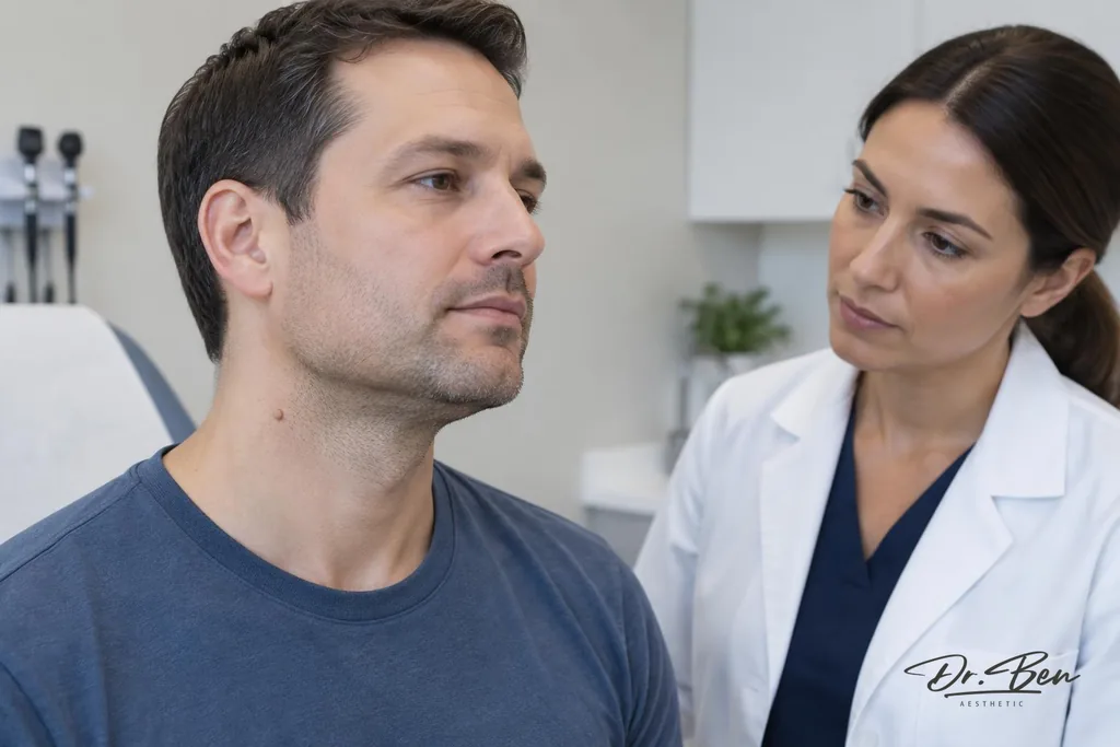
What Is a Benign Skin Lesion?
A benign skin lesion is a non-cancerous growth arising from the skin or the tissues beneath it. Different lesions develop from skin cells, hair follicles, blood vessels, connective tissue or fatty tissue. The term describes the nature of a growth, not one particular appearance or a single diagnosis.
Benign lesions may be flat, raised or felt as a lump under the skin. Their colour, texture and usual location vary, and different types occur at different ages. Some remain unchanged for years, while others become irritated or more noticeable. A lesion that has not been identified should not be assumed benign simply because it is small, painless or resembles a familiar skin tag.
Common Types of Benign Skin Lesions
The following are common examples, but descriptions cannot establish what an individual spot or lump is. Overlap in appearance means that a clinician may need to examine a lesion before offering reassurance or discussing removal.
Skin Tags (Acrochordons)
Skin tags are usually soft growths attached by a narrow stalk. They commonly occur on the neck, in the underarms or groin, beneath the breasts and in other skin folds. Friction may contribute to some tags, and clothing, jewellery or shaving can make them irritated or sore. An atypical stalk-like growth should be examined rather than treated as a skin tag solely on its shape.
Seborrhoeic Keratoses
These common age-related growths may have a waxy, rough or “stuck-on” appearance and range from pale to very dark. They are benign, but some can resemble melanoma or other lesions, particularly when irritated. A new, atypical, bleeding or changing growth should be reviewed before it is labelled a seborrhoeic keratosis.
Benign Melanocytic Moles
Melanocytic moles, or naevi, arise from pigment-producing melanocytes. They may be flat or raised, pigmented or relatively skin-coloured. Most are harmless, but unexpected evolution in size, shape, colour, surface or symptoms warrants medical review. Suspicious or Changing Moles explains changes that deserve attention; a mole should not undergo cosmetic destruction before its diagnosis has been appropriately assessed.
Epidermoid Cysts
Epidermoid cysts are keratin-filled lumps beneath the skin, sometimes with a small central opening called a punctum. They may remain stable, but can rupture or become inflamed and sometimes develop secondary infection. A painful or rapidly enlarging lump needs examination: not every lump with redness or a central mark is a straightforward cyst.
Cherry Angiomas
Cherry angiomas are usually small red or purple vascular bumps that become more common with age. Typical lesions are generally benign. However, an atypical or changing vascular-looking lesion, especially one that repeatedly bleeds, needs assessment rather than reassurance based on colour alone.
Dermatofibromas
Dermatofibromas are firm nodules often found on the arms or legs. Some show a central dimple when the surrounding skin is gently compressed, but that sign is not a reliable home diagnostic test. A nodule that is atypical, rapidly growing or difficult to identify should be examined.
Lipomas
Lipomas are benign fatty-tissue growths that are often soft and mobile beneath the skin. Not all subcutaneous lumps are lipomas. A deep, large, fixed, painful or rapidly growing lump requires evaluation, and examination may lead to further investigation or referral rather than simple reassurance.
Why Do Benign Skin Lesions Develop?
Different lesions have different causes. Age and genetic tendencies influence some growths, while friction may contribute to selected lesions such as skin tags. Other lesions relate to changes in hair follicles, blood vessels, connective tissue or fatty tissue. These are not interchangeable explanations for every lump.
Not all benign growths are caused by ultraviolet exposure, and the presence of several lesions does not necessarily indicate one underlying condition. A consultation considers the type and pattern of lesions and the history of change. It is usually more helpful to establish what a growth is than to assume a cause and try to treat it yourself.
How Can You Tell Whether a Skin Growth Is Benign?
Assessment begins with when the lesion appeared, how it has evolved and whether there is pain, itching, bleeding or irritation. Size, colour, surface texture and, for deeper lumps, mobility and consistency are relevant. Medical history and any previous skin cancer or biopsies can also help guide the examination.
Clinical examination may be enough to identify a typical lesion, but uncertainty sometimes requires further investigation. Dermoscopy uses magnification and illumination to assess skin structures where appropriate; it does not definitively exclude cancer. A biopsy may be needed when the appearance or history raises concern.
Mole & Lesion Assessment provides information about the assessment service. Neither this page nor an online photograph can give a definitive diagnosis for your own lesion. A reassuring resemblance to a picture should not delay review of a new or changing growth.
When Should a Skin Lump or Growth Be Checked?
Seek medical assessment for a new, changing or rapidly growing lesion, an irregular spot, or a growth that bleeds, crusts or does not heal. Pain, inflammation, recurrent irritation and a lump that is difficult to identify are also reasons for review. These signs do not automatically mean cancer, but they should not be dismissed without considering their cause.
An otherwise stable lesion may also be assessed if it catches on jewellery or clothing, is repeatedly injured during shaving or causes cosmetic concern. A cosmetic concern does not mean removal is necessary. The first question remains whether the diagnosis is sufficiently clear and whether observation, investigation or another approach is appropriate.
Seborrhoeic Keratosis Versus Skin Cancer
A seborrhoeic keratosis is benign, but its appearance can overlap with melanoma, basal cell carcinoma or squamous cell carcinoma. Dark pigmentation, a rough surface or an apparently “stuck-on” shape does not provide a definitive diagnosis. An irritated benign keratosis may bleed, but that possibility should not be used to explain away every bleeding lesion.
A changing, atypical or persistently symptomatic growth should be assessed by a clinician. Dermoscopic examination may help, and a biopsy may be appropriate when uncertainty remains. Avoid repeated cosmetic treatment of an undiagnosed lesion or assuming that age-related growths cannot need investigation.
Cysts and Skin Lumps: When Is Further Investigation Needed?
A small, typical cyst may be benign and suitable for observation when its diagnosis is clear. However, deep, large, fixed, rapidly growing, painful, recurrent or difficult-to-identify lumps require a more careful assessment. A lump’s position beneath the skin can make its origin harder to determine from appearance alone.
Examination may lead to imaging, such as ultrasound, or referral where the depth, size or clinical features warrant it. This does not imply that imaging is needed for every cyst or available at every consulting site. The next step depends on the individual lump and the information needed to assess it safely.
Do not repeatedly squeeze or puncture a cyst. This can worsen inflammation, cause trauma and contribute to infection without addressing the cyst lining. A red, swollen or painful lump needs medical review, particularly if redness is spreading or you feel unwell.
What Happens During a Benign Skin Lesion Consultation?
The consultation focuses on identifying the concern and deciding whether anything needs to be done. Bring information about earlier assessments or procedures if available, and describe all symptoms rather than only the appearance you would like changed.
1. Discuss Symptoms and Timeline
Dr Ben asks when you first noticed the growth, whether it has changed and how it affects you. Pain, itching, bleeding, discharge, inflammation and recurrent trauma help guide assessment. Earlier photographs can clarify a timeline, but do not replace examination.
2. Review Relevant Medical History
The discussion includes relevant medical conditions, medicines, allergies and previous procedures. Skin cancer history is considered where relevant, along with factors that may affect bleeding, infection risk or wound healing. Do not stop prescribed medicines on your own in preparation for a possible procedure.
3. Examine the Lesion
Examination considers size, shape, colour, surface, depth, mobility, consistency and signs of inflammation. Dermoscopy may be used where appropriate for the lesion. The clinical findings guide whether a superficial lesion or deeper lump requires further investigation.
4. Explain the Likely Diagnosis and Any Uncertainty
The likely diagnosis is explained along with the limits of the assessment. Where a growth cannot confidently be identified, the plan may include biopsy, imaging or referral rather than immediate removal. Reassurance should be based on the clinical findings, not on an assumption that all common-looking growths are benign.
5. Discuss Options, Risks and Alternatives
Options may include watchful waiting, care for irritation, biopsy, suitable removal or referral. The reasons for an approach, alternatives, costs and relevant risks are discussed before any procedure. There is no obligation to proceed, and treatment at the first appointment should not be assumed.
Does Every Benign Skin Lesion Need Removal?
No. An asymptomatic lesion with an established benign diagnosis can often be observed without intervention. Leaving it in place may avoid a wound, scar or pigment change that is more noticeable than the original growth. Observation is an active choice when the diagnosis and advice about returning for changes are clear.
Removal may be considered for symptoms, recurrent irritation or trauma, a diagnostic need, or an informed elective cosmetic preference. Benign Skin Lesion Services explains the separate procedural service. This concern page does not recommend unnecessary removal or assume that every lesion suitable for assessment is suitable for a procedure.
What Removal Options May Be Discussed?
The technique depends on the diagnosis, size, depth, site and need for tissue examination. These descriptions are general information, not a recommendation or confirmation that each method is offered at both clinics. Actual options and procedural availability must be discussed after examination.
Shave or Snip Removal
Selected superficial lesions may be suitable for shave or snip removal. Whether tissue should be sent for examination depends on the lesion and clinical circumstances. These approaches still create a wound and can cause bleeding, scarring, pigment change or incomplete removal.
Surgical Excision
Excision may be considered according to the lesion’s size, depth and location, particularly where tissue assessment or more complete removal is needed. Sutures and follow-up may be required. The likely scar, healing requirements and whether another setting is more appropriate are considered before proceeding.
Cryotherapy
Cryotherapy may be used for certain appropriately diagnosed benign lesions. It can cause pain, blistering, a burn-like reaction and changes in pigmentation, with variable healing. It is not an appropriate shortcut for destroying a lesion whose diagnosis remains suspicious or uncertain.
Other Destructive or Energy-Based Techniques
Other destructive or energy-based methods may be discussed for selected diagnosed lesions, but should not be used where malignancy is suspected and diagnostic tissue is needed. Cosmetic Mole & Lesion Removal addresses elective removal considerations separately. A preference for a particular technique must not take priority over establishing the diagnosis.
Specialist Management
Large, deep, recurrent, anatomically difficult or diagnostically uncertain lesions may require specialist management. Referral can be the safest way to obtain the necessary assessment or procedure. Mentioning a technique here does not establish that it is suitable for your growth or available at your chosen location.
Why Might a Biopsy or Histopathology Be Needed?
Histopathology is the microscopic examination of tissue by a pathologist. It may be needed when the diagnosis is uncertain, the lesion is atypical or there is concern about malignancy. The result can guide further treatment or follow-up, and the arrangements for receiving results should be explained.
Not every typical benign lesion needs a biopsy, but a suspicious lesion should not be destroyed without considering the diagnostic tissue required. Biopsy & Skin Surgery provides information about tissue sampling and surgical assessment. The appropriate method is selected for the clinical question, rather than treating all lesions in the same way.
Will There Be a Scar After Lesion Removal?
Any excision or destructive procedure can leave a scar. Its appearance depends on the lesion, depth and site, the technique and your wound healing. A previous tendency to hypertrophic or keloid scars is important to discuss, as is a history of pigment changes after injury or procedures.
Scars may be raised, widened or otherwise noticeable, and surrounding skin may become darker or lighter. Healing and the final cosmetic appearance vary between people and body sites. There is no scar-free guarantee; removal should be weighed against observation and the symptoms or concerns caused by the original lesion.
Potential Risks of Skin Lesion Removal
Potential risks include pain, bleeding, bruising, infection, delayed wound healing and scarring. Post-inflammatory hyperpigmentation or hypopigmentation can occur. Allergic reactions and injury to a nerve or nearby tissue are additional considerations depending on the procedure and location.
Incomplete removal, recurrence and a cosmetic result that is disappointing are also possible. The risks are not identical for every technique or lesion. Your medical history, medicines, skin characteristics and healing tendencies help inform the discussion, and informed consent is required before a procedure.
Aftercare may include wound care, sutures, a review appointment or follow-up of pathology results. Clear instructions should explain what is expected and when to seek medical advice for increasing pain, bleeding, redness or other concerns. Costs and alternatives, including no procedure where appropriate, should be understood before proceeding.
Can Benign Skin Lesions Return After Removal?
Some lesions can recur, particularly when removal is incomplete. An epidermoid cyst may return if part of its lining remains. The likelihood depends on the lesion type, technique and clinical circumstances; repeat symptoms should be reassessed rather than automatically treated as the same diagnosis.
New skin tags or seborrhoeic keratoses may also develop elsewhere or near a previously treated area over time. Removing one lesion does not prevent every future growth. No approach can guarantee a recurrence-free result, and a new or changing lesion still warrants appropriate assessment.
When Is Urgent Medical Assessment Important?
Rapidly enlarging, bleeding, painful or unusual lesions need prompt medical assessment. A changing mole or non-healing sore should not be assumed to be a harmless tag or cyst. Describe the change when seeking an appointment, and seek another appropriate medical service if timely assessment is unavailable.
A red, swollen and painful lump may be inflamed or infected. Rapidly spreading redness, fever or feeling systemically unwell warrants urgent medical care rather than waiting for an elective removal visit. Heavy bleeding that does not stop with firm direct pressure also needs urgent care. In a medical emergency call 000.
A broader examination may be appropriate according to your history and findings. Full Skin Checks explains that separate assessment; a concerning individual lesion should not wait until a routine check is due.
Benign Skin Lesion Consultations in Melbourne
Dr Benyamin Mansoori consults at two independent sites. Capstone Medical Centre Southbank is at 216 City Road, Southbank VIC 3006. Cosmetic Avenue Malvern is at 1308 Malvern Road, Malvern VIC 3144. These are independent clinics where Dr Ben sees patients, not clinics owned by him.
Confirm site-specific appointment and procedural availability individually. An assessment does not imply that every removal technique, investigation or specialist service can be provided at that site. The clinical findings determine whether observation, a procedure in an appropriate setting or referral is suitable.
Skin Cancer Fees & Medicare explains relevant fee and eligibility considerations. Benefits depend on the clinical circumstances and applicable criteria; cosmetic-only services generally do not attract Medicare benefits. Costs and any planned investigation or procedure should be clarified before proceeding.
Arrange a Skin Lesion Assessment
A new skin tag, raised mole, cyst or other lump can be assessed to clarify the likely diagnosis and whether further investigation is needed. The outcome may be reassurance and observation, care for symptoms, suitable removal or referral. Diagnosis comes first, and intervention is not necessary for every benign lesion.
Use Book a Skin Lesion Assessment to arrange a consultation and describe any recent changes or symptoms when making the appointment. Do not assume removal will occur at the first visit; suitability, consent, clinical priorities and the relevant clinic’s scheduling all need consideration.
This page is general information, not an individual diagnosis or treatment recommendation. A new, changing, bleeding or otherwise suspicious lesion warrants timely medical evaluation. Text and images on a website cannot establish that your own growth is benign.
Common questions
- What is a benign skin lesion?
- It is a non-cancerous growth of the skin or underlying tissue. Examples include typical skin tags, certain melanocytic naevi, seborrhoeic keratoses, epidermoid cysts and cherry angiomas. A lesion should not be assumed benign from its name or resemblance to an online image; uncertain or changing growths require assessment.
- Are all skin tags harmless?
- Typical skin tags are benign, but not every stalk-like or soft growth is a skin tag. Other lesions can look similar, particularly when irritated or atypical. Have an unusual, changing, bleeding or difficult-to-identify growth examined before considering removal.
- Can a benign mole become cancerous?
- Most benign moles remain harmless. Some melanomas arise in existing moles, while many appear as new lesions rather than developing from an established mole. Unexpected changes in size, shape, colour, surface or symptoms deserve medical review instead of reassurance based on a previous benign appearance.
- What is the difference between a skin tag and a mole?
- A typical skin tag is a soft growth often attached by a narrow stalk; a melanocytic mole arises from pigment-producing cells. Moles can also be raised and skin-coloured, so appearance alone may not distinguish them. Clinical examination helps establish the diagnosis and whether tissue investigation or a particular removal method is appropriate.
- Are seborrhoeic keratoses cancerous?
- Seborrhoeic keratoses are benign growths, not skin cancers. However, they can resemble melanoma and other lesions, and an irritated keratosis can be difficult to identify by appearance alone. A new, atypical, changing or bleeding growth should be examined rather than automatically labelled a keratosis.
- Can an epidermoid cyst become infected?
- Yes. A cyst can become inflamed, for example after rupture, and secondary infection can also occur. Redness or pain does not by itself distinguish inflammation from infection. Spreading redness, fever or feeling unwell requires medical care; avoid repeatedly squeezing or puncturing the lump.
- Does every benign skin lesion need removal?
- No. An asymptomatic lesion with an established benign diagnosis can often be observed. Removal may be considered for symptoms, repeated irritation or trauma, diagnostic reasons or an informed elective preference. The possible wound, scar and other risks should be weighed against leaving the lesion alone.
- Can a benign skin lesion be removed during the first appointment?
- It should not be assumed. The first visit establishes the likely diagnosis and whether observation, investigation, removal or referral is appropriate. A procedure depends on suitability, informed consent, the time and facilities required and the relevant clinic’s scheduling and availability.
- Will skin lesion removal leave a scar?
- Any removal can cause a scar or pigment change, including procedures that do not use sutures. The result varies with depth, location, technique and individual healing, including any tendency to raised or keloid scars. There is no guarantee of a scar-free or cosmetically preferable result.
- Is histopathology always required after removal?
- The need depends on the lesion, the clinical findings and any diagnostic uncertainty or suspicion of cancer. Some typical benign lesions may not require tissue examination, while others do. A suspicious lesion should not be destroyed without considering how an appropriate diagnosis will be obtained.
- Can benign skin lesions return after removal?
- Some can recur, especially if removal is incomplete; an epidermoid cyst may return if lining remains. New skin tags or keratoses can also develop with time. Recurrence depends on the type and method, and no procedure guarantees that a lesion or another growth will never appear again.
- Should I remove a skin tag or mole myself?
- No. Cutting, burning, tying off or using other home removal methods can cause bleeding, infection, scarring and pigment changes. They can also destroy tissue needed to diagnose a lesion that is not what it appears to be. Have the growth assessed before deciding whether removal is appropriate.
Next step
Arrange a consultation
Assessment in person is the only way to work out what is causing a skin concern and what can reasonably be done about it. Fees are explained before anything is arranged.
Medical disclaimer
This page is general information only and is not a diagnosis, a treatment recommendation, or a substitute for individual medical advice. Conditions that look similar can have different causes, and what is suitable for one person may not be suitable for another. If you are unwell, in significant pain, or worried about a rapidly changing symptom, seek medical care promptly. In an emergency call 000.
- Written for patients by
- Dr Benyamin Mansoori, cosmetic physician & skin cancer doctor
- Medically reviewed by
- Dr Benyamin Mansoori (MD, FRACGP, FACCSM), AHPRA MED0002097300
- Last medically reviewed