Medical & Previous Treatment Concerns
Suspicious or Changing Moles & Skin Spots in Melbourne
Dr Benyamin Mansoori provides doctor-led assessment of new or changing moles and skin spots in Southbank and Malvern, Melbourne. Changes in size, shape, colour, texture or symptoms can be reasons to seek medical review, even when a lesion is small or painless.
Most skin lesions are benign, but unusual evolution deserves prompt medical assessment. Melanoma can have an atypical appearance and can occur in hard-to-see areas, including the scalp, back, soles and nail regions. You do not need to be certain that a spot is suspicious before asking for advice.
Clinical examination and dermoscopy where appropriate help guide the next step. Management may involve reassurance, documented monitoring, biopsy, excision or specialist referral. A suspicious lesion should not wait for cosmetic care, and no diagnosis can be made from this website alone.
What Is a Suspicious or Changing Mole?
Common benign moles are collections of melanocytes, the cells that produce pigment in the skin. They may be flat or raised and vary in colour, size and shape. Some have been present since childhood, while others appear later. Normal variation between people is considerable, so a mole does not need to look identical to someone else’s to be benign.
A suspicious mole is one whose appearance, symptoms or pattern of change raises concern during assessment. Evolution in size, shape, colour or texture deserves attention, particularly if it is different from the behaviour of your other moles. Not every change means cancer, but appearance alone cannot reliably settle the question at home.
Mole & Lesion Assessment provides further information about the clinical assessment service. This page focuses on recognising a concern and seeking timely review, rather than identifying a diagnosis from a checklist or photograph.
What Changes in a Mole Should Be Checked?
Arrange medical assessment for a new or unusual spot, a mole that is growing, or a lesion developing asymmetry, an irregular border or several colours. A newly raised lump also deserves review, especially if it is becoming larger over weeks. A spot that stands out from your usual mole pattern may be important even when it is small.
Bleeding without an obvious injury, ulceration, recurrent crusting or a sore that does not heal should be checked. Persistent itching, pain or a change in surface texture can also be relevant. These symptoms have benign causes as well, but should not be dismissed when they occur in a new or evolving lesion.
The absence of pain does not mean a lesion is safe: many skin cancers cause little or no discomfort at first. If you cannot explain a change, or someone notices an unusual spot on your back or scalp, seek examination rather than waiting for symptoms to become more obvious.
The ABCDE Guide to Checking Moles
ABCDE is an educational guide to features that may warrant medical assessment. It is not a diagnostic test, and a lesion can need investigation without meeting every letter. Comparing a spot with its earlier appearance and with your other moles is often more useful than focusing on one feature.
A — Asymmetry
One half of a mole may look different from the other in shape, colour or internal pattern. Some benign moles are not perfectly symmetrical, but new or increasing asymmetry should be reviewed.
B — Border
An irregular, notched, uneven or poorly defined edge can be concerning. A border that has changed is worth bringing to your doctor’s attention even if the overall lesion remains small.
C — Colour
Several shades within one lesion, or a new area of black, brown, red, white or blue, may warrant examination. Melanoma is not always dark, so a pink or skin-coloured spot should not be ignored simply because it lacks brown pigment.
D — Diameter
A diameter greater than about 6 mm is a common prompt for checking a mole, not a threshold below which melanoma is excluded. Melanomas can be smaller than 6 mm, and increasing size matters at any starting diameter.
E — Evolution
Evolution means change over time, including size, shape, colour, elevation, texture or symptoms such as bleeding or itching. A clearly changing lesion deserves assessment even if its border looks regular or its colour seems uniform.
Melanomas That Do Not Follow the ABCDE Pattern
Some melanomas contain little visible pigment and may appear pink, red or skin-coloured. These amelanotic melanomas can resemble benign growths or other skin conditions. A persistent or evolving non-pigmented lesion still needs assessment; colour is only one part of the clinical picture.
Some nodular melanomas grow rapidly as a raised lump and may look relatively uniform or symmetrical. Waiting for the classic irregular, multicoloured appearance can delay assessment of a lesion that requires investigation.
The EFG Warning Features
EFG refers to Elevated, Firm and Growing. A raised, firm lesion that is enlarging, particularly over weeks, should be examined promptly. These features are suggestive rather than specific: benign lesions can also be raised or firm, and their absence does not exclude melanoma. Do not use EFG to reassure yourself about a spot that is otherwise changing.
The Ugly Duckling Sign
Many people have a recognisable pattern among their own moles. An “ugly duckling” is a spot that looks noticeably different from that pattern: it might be darker, lighter, larger, smaller or shaped differently from the surrounding moles.
The comparison is with your own skin, not a photograph of someone else’s melanoma. A standout lesion should be reviewed even when it does not show all of the ABCDE features. If you have very few moles, a single new or unusual lesion can still merit attention.
Can a Suspicious Skin Spot Be Something Other Than Melanoma?
Yes. Several benign conditions and other skin cancers can look similar. Clinical examination helps determine which possibilities need consideration, and tissue examination may be needed when the diagnosis remains uncertain. The following descriptions are not a way to diagnose a spot yourself.
Benign Moles
Benign moles can be flat, raised, lightly pigmented or darker and may change gradually during life. A familiar appearance may be reassuring in context, but a new or unexpected change still deserves review. Benign Skin Lesions explains other common non-cancerous growths without assuming that every new lump is harmless.
Seborrhoeic Keratoses
These common benign growths may look waxy, rough or “stuck on” and can be brown or nearly black. Irritation can cause crusting or bleeding. Because some resemble other lesions, an unfamiliar or changing growth should be assessed before it is treated as a seborrhoeic keratosis.
Solar Lentigines
Solar lentigines are flat areas of pigmentation associated with sun exposure. They commonly occur on the face, hands and other exposed skin. An irregular or evolving pigmented patch should not automatically be labelled a sun spot; examination may be needed to distinguish it from other causes.
Basal Cell Carcinoma
Basal cell carcinoma may appear as a pearly or pink bump, a persistent patch, or a sore that repeatedly bleeds or fails to heal. It does not always resemble a mole. Although its behaviour differs from melanoma, assessment and an appropriate management plan remain important.
Squamous Cell Carcinoma
Squamous cell carcinoma can appear as a scaly, thickening or crusted lesion, sometimes with tenderness, ulceration or growth. These appearances overlap with other conditions, so a persistent or enlarging spot needs medical review rather than repeated cosmetic treatment.
Melanoma
Melanoma can be pigmented or non-pigmented, flat or raised, and may arise in a pre-existing mole or as a new lesion. No single visible feature confirms or excludes it. Where a skin cancer is diagnosed, Skin Cancer Treatment outlines subsequent management considerations; the immediate priority for an undiagnosed changing lesion is establishing what it is.
Who May Have an Increased Risk of Skin Cancer?
Relevant factors include a personal history of skin cancer, a family history of melanoma, substantial ultraviolet exposure or sunburns, skin that burns easily, numerous or atypical moles, and previous solarium use. Immunosuppression can increase risk, and increasing age is relevant to some skin cancers.
Risk is individual: having one of these factors does not mean a changing spot is cancer, and having none does not make it safe to ignore. Tell your doctor about medicines or conditions that affect your immune system and any previous biopsies or skin cancer diagnoses.
Skin cancer can affect people of all skin tones and can occur at sites with little sun exposure, including palms, soles and nail areas. Assessment should respond to the lesion itself as well as the person’s background risk.
How Should You Check Your Skin at Home?
Become familiar with your usual skin appearance in good light. Look at your face, ears, scalp, neck, torso, back, arms and legs, including between fingers and toes, the soles of your feet and around or beneath nails. Hard-to-see areas matter; a mirror or help from a partner may make them easier to inspect.
Notice new spots and changes rather than trying to assign a diagnosis. If you already have photographs, they may help you describe when a change occurred, but a photograph is not a substitute for examination. Do not postpone an appointment to collect more pictures of a lesion that is growing or otherwise concerning.
Professional skin check frequency should be individualised according to history, risk and examination findings. There is no single automatic interval suitable for everyone. Seek review of a changing lesion between planned checks rather than assuming it can wait until the next routine appointment.
What Happens During a Suspicious Mole Assessment?
The consultation is directed at the spot that concerns you and the clinical context around it. Mention all lesions you are worried about when arranging and attending the appointment so the scope of examination can be discussed.
- Your concern and timeline: Dr Ben asks when the spot was first noticed, how it has changed and whether there has been bleeding, itching, pain or a non-healing surface. Earlier photographs or records can be useful if available.
- Personal and family risk history: the discussion includes previous skin cancers or biopsies, family melanoma history, UV exposure, sunburns and relevant medical conditions or medicines.
- Clinical examination and comparison: the lesion’s site, shape, colour, surface and other features are examined, with comparison to other lesions where appropriate. A broader examination may be discussed according to the findings and your risk.
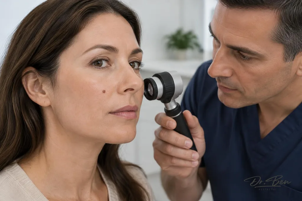
- Dermoscopic assessment where appropriate: a trained clinician may use a dermatoscope to examine structures not readily visible to the naked eye. The findings are considered alongside the history and clinical appearance.
- An individual management decision: this may be reassurance, documented monitoring or photography, biopsy, complete excision or specialist referral. The reason for the recommendation, follow-up arrangements and what changes should prompt earlier review are explained.
Monitoring is not suitable for every lesion. A spot with high clinical suspicion may require prompt tissue diagnosis rather than a period of observation. Consultation and procedure arrangements depend on the lesion, clinical needs and availability; a procedure is not automatically undertaken at the first visit.
What Is Dermoscopy?
Dermoscopy is a non-invasive examination using magnification and illumination to assess skin structures and patterns that are not readily seen with the naked eye. A clinician trained in its use interprets these features together with your history and the appearance of the lesion.
It can improve clinical assessment, but it does not definitively exclude malignancy or replace histopathology when suspicion remains. Histopathology means examination of tissue by a pathologist, and may be necessary to establish the diagnosis.
Website images, phone photographs and automated or AI-based assessments cannot substitute for appropriate professional investigation. Do not let a reassuring app result delay medical examination of a new, evolving or otherwise unusual spot.
Monitoring, Biopsy or Excision: How Is the Decision Made?
The decision depends on the degree of suspicion, lesion type and location, your medical circumstances and the information needed to make a diagnosis. Your doctor should explain why an approach is recommended and how the result or follow-up will be communicated.
Monitoring
Documented monitoring may be appropriate for selected lesions when the assessed risk supports observation. It should have a planned interval, a method of comparison where useful and clear instructions for returning sooner if changes occur. High suspicion generally calls for prompt histological assessment, not watchful waiting.
Biopsy
A biopsy provides tissue for examination by a pathologist. The technique is chosen for the specific lesion and clinical question. For suspected melanoma, an appropriate full-thickness excisional biopsy of the lesion is generally preferred when feasible; difficult locations, lesion size or other circumstances may require a different approach or referral. Biopsy & Skin Surgery provides information about these procedures and their practical considerations.
Excision
Excision removes a lesion surgically and allows the tissue to be examined. The pathology result determines whether further treatment or follow-up is needed; initial removal is not a promise that all subsequent care is complete. Scarring, bleeding, infection and healing considerations are discussed as part of consent.
Specialist Referral
Referral may be appropriate where there is diagnostic uncertainty, a high-risk lesion, a difficult anatomical site or a need for more complex management. A confirmed malignancy may require additional treatment planning. Referral is part of responsible care, not a reason to delay investigation while pursuing a cosmetic solution.
Focused Mole Assessment Versus a Full Skin Check
A focused appointment addresses a particular new, changing or concerning lesion. A full skin check is a broader examination intended to assess the skin more widely, with its scope guided by your history, risk and consent. A focused lesion assessment should not be assumed to include a complete examination of every skin area.
Full Skin Checks explains the broader assessment service. If you have an evolving spot, do not wait for a routine full check before seeking advice. Your doctor can assess the immediate concern and discuss whether a wider examination or follow-up is also appropriate.
Should a Suspicious Mole Be Removed for Cosmetic Reasons?
The priority is establishing the diagnosis, not improving the appearance. A suspicious lesion should not undergo cosmetic laser treatment or another destructive method that prevents appropriate tissue examination. Destroying a lesion without histology can obscure the diagnosis and delay necessary care.
Cosmetic Mole & Lesion Removal is a separate discussion for lesions assessed as suitable for elective removal. Even a likely benign lesion requires appropriate assessment, consent and discussion of scarring, recurrence and whether histology is indicated. Do not choose a cosmetic procedure as a shortcut around investigating a cancer concern.
When Should You Arrange Medical Assessment?
Seek prompt medical assessment for a new or evolving lesion, rapid growth, recurrent bleeding or crusting, ulceration, a non-healing spot or an ugly duckling lesion. A changing spot on a foot or around a nail, including a new or changing pigmented nail band, also deserves examination. These changes do not establish a diagnosis, but should not be ignored.
Do not delay while waiting for a cosmetic appointment, repeated self-tests or an app result. When arranging care, describe any rapid growth, bleeding or other concerning change so the service can advise on timing. If a timely appointment is unavailable, seek another GP or appropriate medical service rather than leaving the lesion unassessed.
If bleeding is heavy or will not stop with firm direct pressure, seek urgent medical care. For a medical emergency call 000. This emergency advice is separate from the need for prompt examination of a changing lesion that is otherwise not causing an acute emergency.
Can Skin Cancer Be Prevented?
Not all skin cancers can be prevented, but reducing ultraviolet exposure lowers avoidable risk. Use broad-spectrum sunscreen as directed, protective clothing, a hat, shade and sunglasses, and avoid solaria. Sunscreen is one part of sun protection, not permission to spend longer in the sun.
Skin awareness and appropriate medical review remain important even when you practise sun protection. Skin cancer can occur in all skin tones and at less sun-exposed sites. Advice about professional checks should reflect your individual risk rather than relying on a universal schedule or assuming prevention eliminates the need to investigate a change.
Suspicious Mole Consultations in Melbourne
Dr Benyamin Mansoori consults at two independent sites in Melbourne. Capstone Medical Centre Southbank is at 216 City Road, Southbank VIC 3006. Cosmetic Avenue Malvern is at 1308 Malvern Road, Malvern VIC 3144. These are independent clinics where Dr Ben sees patients, not clinics owned by Dr Ben.
Appointment and procedural availability must be confirmed individually with the relevant service. The location of an appointment does not mean every procedure or specialist service is available there. If the lesion requires care beyond the consultation setting, biopsy arrangements or referral can be discussed according to the clinical findings.
Skin Cancer Fees & Medicare explains fee and eligibility considerations. Medicare benefits depend on the clinical circumstances and applicable criteria; they are not guaranteed for every assessment or procedure. Fees and any planned investigation should be clarified before proceeding.
Arrange a Mole and Skin Lesion Assessment
A new, changing or unusual mole or skin spot warrants prompt examination rather than a cosmetic decision based on its appearance. Assessment may include clinical examination and dermoscopy where appropriate, with monitoring, biopsy, excision or referral determined by the level of concern and your circumstances.
To arrange care, use Book a Mole Assessment and describe the lesion and any recent changes when making your appointment. If access is delayed and the spot is rapidly growing, bleeding or otherwise concerning, seek another appropriate medical service for timely review.
This page provides general information only. A suspicious mole cannot be diagnosed or ruled out from website text or an image, and this information does not replace individual medical advice. Prompt in-person examination is the appropriate next step when a lesion is concerning.
Common questions
- Does a changing mole always mean skin cancer?
- No. Moles and other benign lesions can change, including through irritation or normal changes over time. However, benign and malignant lesions may look similar, so a new or unexpected change deserves prompt medical assessment rather than reassurance based on appearance alone.
- What are the ABCDE signs of melanoma?
- A is asymmetry, B is an irregular border, C is colour variation, D is diameter or increasing size, and E is evolution in appearance or symptoms. These features help identify lesions that need review, but are not diagnostic. Some melanomas do not follow the ABCDE pattern, and a lesion need not show every feature to warrant examination.
- Can melanoma be smaller than 6 mm?
- Yes. Melanoma can be smaller than 6 mm, particularly when first noticed. The commonly used diameter guide is a prompt for assessment, not a safe-size cut-off. Change, unusual appearance and other clinical features matter regardless of the measurement.
- Can melanoma be pink or skin-coloured?
- Yes. Amelanotic melanoma has little visible pigment and may appear pink, red or skin-coloured. A persistent or changing non-pigmented lesion should be assessed, especially if it is growing, bleeding or not healing. Lack of dark pigment does not exclude melanoma.
- What is the difference between a normal mole and a suspicious mole?
- Many benign moles are stable and fit a person’s usual mole pattern, while an evolving, unusual or symptomatic lesion may raise concern. There is considerable overlap in appearance, and no home comparison reliably establishes the diagnosis. Clinical examination, sometimes with dermoscopy or tissue investigation, is needed to assess a suspicious spot.
- What is the ugly duckling sign?
- It describes a lesion that looks noticeably different from your other moles, whether in colour, shape, size or overall pattern. The comparison is with your own skin. A standout lesion should be reviewed even if it does not meet all the ABCDE features.
- Does dermoscopy confirm whether a mole is cancerous?
- Not on its own. Dermoscopy helps a trained clinician assess structures and patterns beneath the visible surface, but cannot provide a definitive guarantee that a lesion is benign. When cancer is suspected or uncertainty requires investigation, tissue examination by a pathologist may be necessary.
- Will every suspicious mole need a biopsy?
- The decision depends on the clinical assessment and degree of suspicion. Selected lesions may be suitable for documented follow-up, while others need prompt biopsy, excision or specialist referral. Monitoring should have clear arrangements and is not appropriate for every concerning lesion.
- Can a suspicious mole be removed with a laser?
- Cosmetic laser destruction is inappropriate when cancer is suspected because tissue may be needed for diagnosis. The lesion should be assessed first and investigated using an appropriate method. Elective removal of a lesion judged benign is a separate consent discussion, not an alternative to evaluating a suspicious spot.
- Is a focused mole assessment the same as a full skin check?
- No. A focused assessment addresses a particular lesion or concern, while a full skin check examines the skin more broadly according to history, risk and consent. Discuss the intended scope when arranging care. Do not wait for a routine full check before seeking assessment of a clearly changing lesion.
- Can skin cancer occur in people with darker skin?
- Yes. Skin cancer can affect people of all skin tones, including at sites such as the palms, soles and nails. Lower background risk does not make a changing lesion harmless. New or evolving spots should be assessed on their own clinical features.
- How soon should I see a doctor about a changing mole?
- Arrange prompt medical assessment rather than waiting for a cosmetic visit or your next routine check. Tell the service if the lesion is rapidly growing, bleeding, crusting, ulcerated or not healing so timing can be advised. If a timely appointment is unavailable, seek another GP or appropriate medical service instead of postponing care.
Next step
Arrange a consultation
Assessment in person is the only way to work out what is causing a skin concern and what can reasonably be done about it. Fees are explained before anything is arranged.
Medical disclaimer
This page is general information only and is not a diagnosis, a treatment recommendation, or a substitute for individual medical advice. Conditions that look similar can have different causes, and what is suitable for one person may not be suitable for another. If you are unwell, in significant pain, or worried about a rapidly changing symptom, seek medical care promptly. In an emergency call 000.
- Written for patients by
- Dr Benyamin Mansoori, cosmetic physician & skin cancer doctor
- Medically reviewed by
- Dr Benyamin Mansoori (MD, FRACGP, FACCSM), AHPRA MED0002097300
- Last medically reviewed