Skin Cancer
Skin Cancer Checks in Melbourne: When to Get Checked and What to Expect
- By
- Dr Benyamin Mansoori
- Published
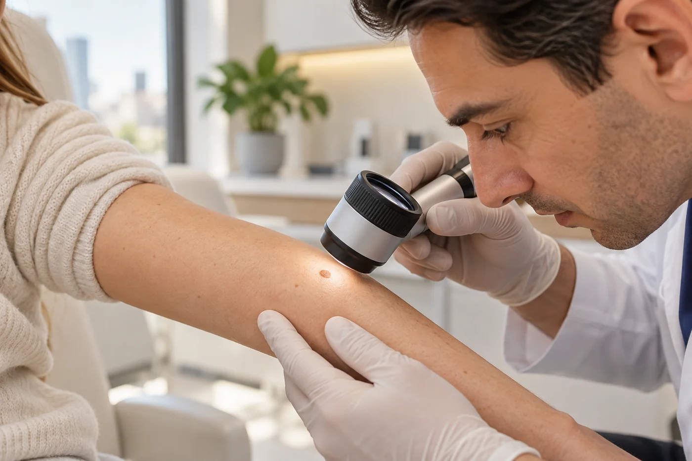
Skin cancer checks in Melbourne: learn warning signs, what dermoscopy involves, and when professional skin checks may be appropriate for your risk.
Australia has one of the world's highest rates of melanoma. The Australian Institute of Health and Welfare estimated approximately 17,400 new melanoma cases in Australia in 2025. This figure is an estimate of melanoma cases, not a count of all skin cancers. [1]
A new spot, a changing mole or a sore that does not heal can be worrying. Many skin changes are harmless, but some need prompt medical assessment. Understanding the warning signs and what happens during a skin check can help you make informed decisions.
This guide explains the main types of skin cancer, the ABCDE and EFG warning signs, dermoscopy, and how the need for professional skin checks varies with individual risk. It is intended for patients in Melbourne and across Australia.
What Is a Skin Cancer Check?
A skin cancer check is a clinical examination to identify skin lesions that may require further assessment. A consultation may focus on one concerning spot or involve a full-body examination, depending on your symptoms, risk factors and needs. The doctor asks about changes you have noticed and may use a handheld instrument called a dermatoscope to examine selected lesions more closely. [2,3]
A skin check cannot guarantee detection of every cancer. Continue to pay attention to your skin between appointments, and seek assessment for a new or changing spot even if your previous examination was reassuring.
The Three Main Types of Skin Cancer
Basal Cell Carcinoma (BCC)
BCC is the most common type of skin cancer in Australia. It may look like a pearly or pink bump, a pale or scaly patch, or a sore that repeatedly crusts or fails to heal. BCC often develops on sun-exposed skin. It usually grows slowly and rarely spreads to distant sites, but can damage surrounding tissue when untreated. [3,4]
Squamous Cell Carcinoma (SCC)
SCC may present as a thickened scaly spot, a tender or growing lump, or a persistent crusted or ulcerated lesion. It often develops on skin with cumulative ultraviolet (UV) exposure. Some SCCs can grow deeply or spread, making timely assessment important. [3,4]
Melanoma
Melanoma develops from pigment-producing melanocytes and can spread to other parts of the body. It may arise as a new spot or within an existing mole. Some melanomas are brown or black, while others are pink, red or relatively unpigmented. Melanoma can occur in places not usually exposed to sunlight. [4,5]
Not every suspicious skin lesion is cancer, and appearance alone cannot always determine the diagnosis.
How to Recognise a Suspicious Mole: The ABCDE Guide
The ABCDE rule is a practical guide to changes that deserve medical review. It is not a diagnostic test, and melanoma can occur without all five features. [4,5]
- A — Asymmetry: one half of the spot differs from the other.
- B — Border: irregular, notched or blurred edges.
- C — Colour: uneven colour or several shades within the same lesion.
- D — Diameter: the lesion is getting larger; melanoma can also be smaller than 6 mm.
- E — Evolving: a change in size, shape, colour, elevation or symptoms over time.
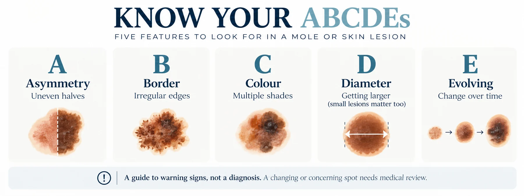
Also be alert to an “ugly duckling” spot that looks noticeably different from your other moles. New bleeding, crusting or persistent irritation can be important even when the lesion does not fit the ABCDE pattern. [4]
Additional Warning Signs: EFG
Some rapidly growing melanomas, including nodular melanoma, do not follow the familiar ABCDE pattern. Cancer Council NSW also describes the EFG features: Elevated (raised), Firm (firm to touch) and Growing (growing quickly). Such lesions may be skin-coloured, pink, red, brown or black. [5]
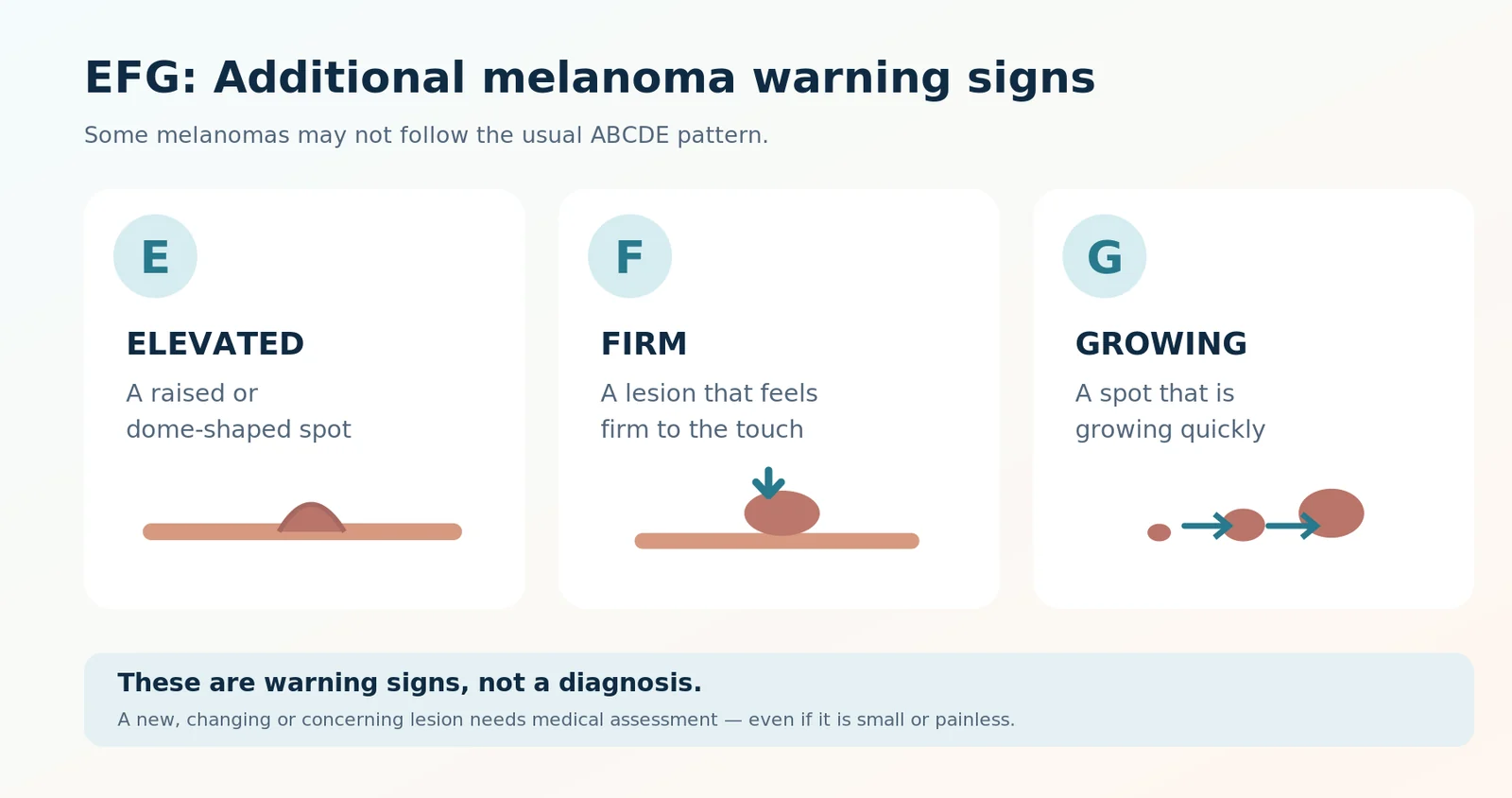
Do not wait for a spot to become dark, large or painful before having it examined.
Who May Benefit from a Professional Skin Check?
Anyone with a new, changing or concerning lesion should seek medical assessment, regardless of risk category. Factors associated with higher skin cancer risk include: [2,6]
- A personal history of melanoma or another skin cancer.
- A first-degree relative with melanoma.
- Numerous or atypical moles.
- Fair skin, a tendency to burn, or substantial previous UV exposure.
- Significant occupational or recreational sun exposure.
- Immunosuppression, including some medical treatments.
Skin cancers can occur in all skin tones. People with darker skin can also develop suspicious lesions, including on the palms, soles or beneath the nails; a concerning change deserves attention regardless of skin colour. [4]
How Often Should You Have a Skin Cancer Check?
There is no universal recommendation for every Australian adult to have an annual full-body skin check. Cancer Council Australia does not recommend population-wide screening because high-quality evidence of a reduction in deaths across the general population is insufficient. This is different from assessing a person who already has a suspicious lesion. [2]
The RACGP's Guidelines for Preventive Activities in General Practice (Red Book, 10th edition) offers a risk-based approach: [6]
- Average or below-average risk: routine screening examinations are generally not recommended when there is no clinical concern.
- Above-average risk: an opportunistic clinical examination may be appropriate, usually no more often than annually.
- High risk: regular clinician skin examinations are recommended, generally at least annually.
- Very high risk of a new primary melanoma: six-monthly full-body examinations supported by dermoscopy and total-body photography may be appropriate under an individualised programme.
Your clinician should determine which category applies to you. These intervals are not instructions to delay assessment: a concerning change should be assessed promptly at any time.
What Happens During a Skin Cancer Check?
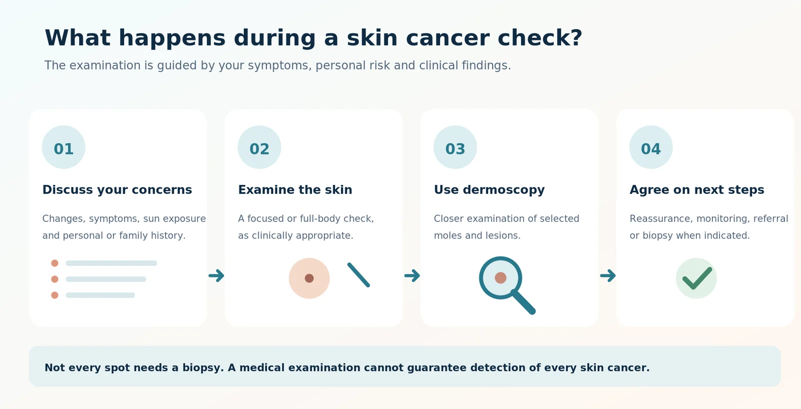
1. Medical History and Risk Assessment
Your doctor may ask about skin changes, previous skin cancer, family history, immune conditions and sun exposure. Mention any lesion that is new, growing, bleeding, crusting or otherwise worrying you.
2. Examination of the Skin
The doctor examines the relevant skin areas. If a full-body check is indicated, the examination may include the scalp, back, ears, hands, feet and other easily overlooked sites. The examination should respect your privacy, preferences and consent.
3. Dermoscopy
A dermatoscope magnifies the skin and reveals patterns and structures not easily visible to the naked eye. A Cochrane systematic review found that dermoscopy, particularly when used by experienced clinicians, was more accurate for melanoma detection than visual inspection alone. However, included studies had important limitations, evidence from primary care was relatively limited, and no technique detects every melanoma. [7,8]
Dermoscopy supports clinical judgement; it does not replace pathology when a biopsy is indicated.
4. Results and a Personalised Plan
Depending on the findings, the clinician may advise reassurance, monitoring, photography, biopsy, excision or referral. Not every spot requires removal. The recommendation should reflect the lesion, risk factors, diagnostic uncertainty and your preferences. [3]
What If a Suspicious Lesion Is Found?
A suspicious appearance does not automatically mean cancer. When necessary, a clinician may recommend a skin biopsy, in which tissue is collected and examined under a microscope by a pathologist. The choice of biopsy technique depends on the suspected diagnosis, the site and size of the lesion, and other clinical factors. A lesion suspicious for melanoma requires an appropriate diagnostic approach. [3,9]
Biopsy or excision can involve discomfort, bleeding, infection, scarring and, occasionally, further procedures. Your clinician should explain the reasons, alternatives and relevant risks before obtaining consent. Pathology results then help guide any further treatment or follow-up.
For more information, see Mole and Lesion Assessment and Biopsy and Skin Surgery.
How to Check Your Skin at Home
Become familiar with your skin so that new or changing spots are easier to notice. Check in good light, use a mirror for difficult-to-see areas, and ask a trusted person for help if needed. Remember the scalp, back, ears, hands, feet, between the toes, soles and nails. [4]
There is no single self-check interval suitable for everyone. Your doctor can advise an approach based on your individual risk. Self-examination supports skin awareness but cannot rule out skin cancer or replace assessment of a worrying lesion.
Do not rely on a smartphone application or online photograph to declare a suspicious spot benign. [4]
Reducing Your Risk: Sun Protection in Australia
UV radiation is a major preventable cause of skin cancer. Cancer Council Australia advises using all five SunSmart measures when the UV Index is 3 or above: [10]
- Slip on protective clothing.
- Slop on broad-spectrum, water-resistant SPF50 or SPF50+ sunscreen.
- Slap on a broad-brimmed hat.
- Seek shade.
- Slide on sunglasses that meet Australian Standards.
Apply sunscreen generously around 20 minutes before UV exposure and reapply at least every two hours outdoors, as well as after swimming, sweating or towel-drying. Sunscreen supplements other protective measures and is not a reason to stay outside longer. [10]
Skin Cancer Assessment with Dr Ben Aesthetic in Melbourne
Dr Ben Aesthetic provides doctor-led skin checks, assessment of concerning moles and dermoscopy in Melbourne. An appointment can help clarify individual risk, assess a lesion and discuss reasonable next steps where further investigation is appropriate.
Read more about Full Skin Checks, Dermoscopy, and Preparing for a Skin Check. For details about consultation charges and individual eligibility for rebates, see Skin Cancer Fees and Medicare.
If you notice a suspicious or changing lesion, arrange medical assessment rather than waiting for your next routine appointment.
Frequently Asked Questions
Does a skin cancer check hurt?
A visual examination and dermoscopy are non-invasive and are generally not painful. If a biopsy is recommended, anaesthesia, possible discomfort and procedure risks should be explained beforehand.
Does every Australian need an annual skin check?
No. Routine annual screening of all average-risk people without symptoms is not recommended. The appropriate approach depends on individual risk, previous findings and whether a suspicious lesion is present. [2,6]
Can a small or pink spot be melanoma?
Yes. Melanomas may be smaller than 6 mm or pink, red or relatively unpigmented. A lesion that changes or grows quickly requires assessment. [4,5]
Does dermoscopy confirm melanoma?
No. Dermoscopy can improve clinical assessment but is not perfectly accurate. A biopsy with histopathological examination may be needed for a definitive diagnosis. [3,7]
Will a suspicious mole always be removed?
No. Some lesions can be monitored or reviewed, while others warrant biopsy, excision or specialist assessment. Your clinician will explain the reasons for the proposed plan.
Are skin checks covered by Medicare?
Some eligible medical consultations and clinically indicated procedures may attract a Medicare rebate, but this depends on eligibility and the applicable item requirements. Ask the clinic to confirm fees and possible out-of-pocket costs.
Should I wait until my scheduled skin check if a spot is changing?
No. Arrange prompt medical review for a growing, changing, bleeding or non-healing lesion rather than waiting for a routine appointment.
Sources and further reading
- Australian Institute of Health and Welfare. Cancer data in Australia: Melanoma of the skin in Australia. Updated 2026.
- Cancer Council Australia. Early Detection of Skin Cancer: Position Statement. January 2019.
- Cancer Council Australia. Diagnosing skin cancer. Updated 21 September 2026.
- Cancer Council Australia. Check for signs of skin cancer.
- Cancer Council NSW. Signs and symptoms of melanoma. Updated March 2025.
- Royal Australian College of General Practitioners. Guidelines for Preventive Activities in General Practice, 10th edition, skin cancer recommendations (2024).
- Dinnes J, Deeks JJ, Chuchu N, et al. Dermoscopy, with and without visual inspection, for diagnosing melanoma in adults. Cochrane Database Syst Rev. 2018;12:CD011902.
- Dinnes J, Deeks JJ, Grainge MJ, et al. Visual inspection for diagnosing cutaneous melanoma in adults. Cochrane Database Syst Rev. 2018;12:CD013194.
- Cancer Council Australia and Melanoma Institute Australia. Clinical practice guidelines for the diagnosis and management of melanoma.
- Cancer Council Australia. Preventing skin cancer and Advice on sunscreen use.
Related pages
Medical disclaimer
This content is general information only and is not a diagnosis, a treatment recommendation or a substitute for personal medical advice. If you notice a new or changing spot, bleeding or a non-healing sore, or you are worried about a symptom, arrange a medical assessment. In an emergency call 000.